C&B Metabond

499.07

C&B Metabond R&D cement system for in vivo brain research. Bonds metals to skull/bone without glue. Developed with European neuroscientists for lab use.

Find your local distributor or contact us.

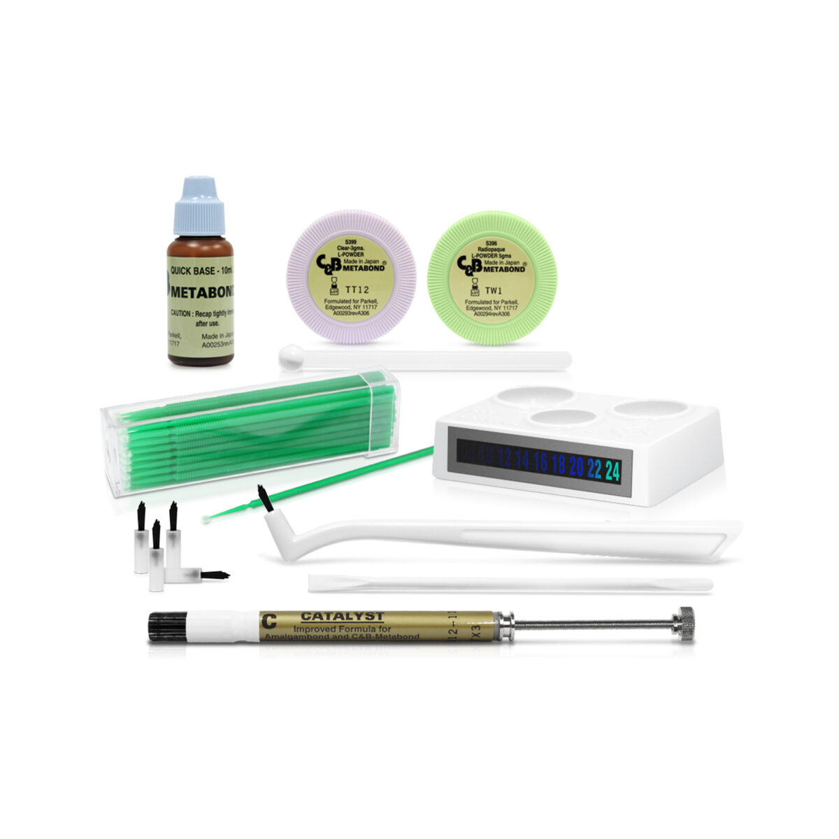
Includes, “B” Quick Base (10 ml), Catalyst (0.7 ml), Radiopaque L-Powder (5 gm), Clear L-Powder (3 gm), Adjustable Applicator Brushes, Disposable Applicator Brush Tips, Ceramic Mixing Dish with Thermometer & Mixing & Application Accessory Kit.

Description

C&B Metabond R&D

The C&B Metabond R&D Dental Cement stands as the gold standard for adhesive bonding in neuroscience and brain research. Nearly all major U.S. and international institutions—including Oxford University, the NIH, Karolinska Institutet and the Allen Institute—use and rely on this adhesive luting cement. It is engineered to bond any substrate to skull or bone, especially metals, with outstanding strength and stability.

In contrast to acrylic systems, which often require glue layers or complicated handling, C&B Metabond R&D offers a cleaner, stronger, and more direct bond—without the need for additional adhesives. The R&D kit was developed in collaboration with European neuroscientists to meet the specific needs of in vivo research. As a result, this version includes only the components necessary for experimentation, avoiding all unnecessary materials and eliminating clinical waste.

For Research Purposes Only

Please note: C&B Metabond Mouse Surgery Adhesive is not CE-marked and is not approved for dental use in Europe. It is intended exclusively for non-human research applications.

What’s Included in the C&B Metabond R&D a dental adhesive for rodent surgery (Item # S380-RD):

  • (1) “B” Quick Base (10 ml)

  • (1) Catalyst (0.7 ml)

  • (1) Radiopaque L-Powder (5 gm)

  • (1) Clear L-Powder (3 gm)

  • (50) Adjustable Applicator Brushes

  • (100) Disposable Applicator Brush Tips

  • (1) Ceramic Mixing Dish with Thermometer

  • (1) Mixing & Application Accessory Kit

Refills & Accessories (available separately)

You can reorder all components individually, including brush tips, powders, dentin activator, and mixing kits. Parkell Europe ships these articles worldwide, orders typically deliver in approximately one to two weeks. [Click here to purchase refills & accessories separately.]

Referenced in Leading Scientific Protocols

C&B Metabond has appeared in dozens of published neuroscience studies, including research conducted by the Allen Institute, International Brain Laboratory, and Oxford University. Scientists cite it consistently for its stability, reliability, and clean handling during cranial implant surgeries and long-term recordings.

Order C&B Metabond® R&D for high-performance adhesive stability in non-dental, research-based neurobiology applications worldwide.

A selection of article links related to the use of C&B Metabond R&D as a dental adhesive for rodent surgery can be found below:

Allen Institute for the Brain : A Standardized Head-Fixation System for Performing Large-Scale, In Vivo Physiological Recordings in Mice.

International Brain Laboratory Protocol: 2020: Appendix 1: IBL protocol for headbar implant surgery in mice, v4.

Barkus et al. (2022): Refinements to rodent head fixation and fluid/food control for neuroscience.

Coelho-Santos et al. (2022): Reinforced thinned-skull window for repeated imaging of the neonatal mouse brain.

Silasi et al. (2016): Intact skull chronic windows for mesoscopic wide-field imaging in awake mice.

Goldey et al. (2014): Long-term imaging in awake mice using removable cranial windows.

Wang et al (2022): Protocol for recording epileptiform discharges of EEG and behavioral seizures in freely moving mice. STAR Protocol.

Order C&B Metabond® R&D for high-performance adhesive stability in non-dental, research-based neurobiology applications across Europe.
Meet us in Barcelona at the FENS Forum (Federation of European Neuroscience Societies),  where C&B Metabond R&D will be available.

Videos

video-thumbnail

C&B Metabond R&D- How to Use Video

Information

Description
File name
Language

Scientific Literature

Explore published neuroscience research involving standardized head-fixation systems  and other in vivo studies in mice.

A standardized head-fixation system for performing large-scale, in vivo physiological recordings in mice
Groblewski et al. · 2020 · Journal of Neuroscience Methods

Groblewski et al. (2020), researchers at the Allen Institute for Brain Science, developed a standardized head-fixation system for high-throughput optical physiology, electrophysiology, and behavioral experiments in awake mice.

The system combined a titanium headframe, reproducible clamping system, surgical tooling, and a shared coordinate framework. During the surgical procedure, C&B Metabond® was used to affix the titanium headframe to the mouse skull before subsequent positioning, craniotomy, and recording procedures.

Key findings
  • The complete head-fixation system was designed to provide reproducible positioning and rigid fixation across multiple experimental platforms.
  • When used with the clamping system, the titanium headframe showed less than 4 μm of deflection in simulations, bench testing, and in vivo testing.
  • The headframe was reported to be well tolerated by the animals and allowed technicians to perform rapid and consistent head fixation.
  • C&B Metabond formed part of the standardized surgical workflow by securing the titanium headframe to the skull.

We developed a headframe (and associated surgical tooling and clamping system) that met all of our pre-defined requirements pertaining to registration, rigidity, compatibility with our experimental systems, animal health, ease of handling, and future adaptability.

The headframe design incorporated features that created three mutually intersecting perpendicular datum planes and, when used in conjunction with our clamping system, showed less than 4 μm of deflection in our simulations, bench tests, and in vivo tests.

Study context

This was an in vivo methods and engineering study involving mice. The reported rigidity and deflection results apply to the complete headframe, tooling, and clamping system, not to C&B Metabond alone. The study documents the use of C&B Metabond to affix the titanium headframe.

Reference

Groblewski, P. A., Sullivan, D., Lecoq, J., de Vries, S. E. J., Caldejon, S., L’Heureux, Q., Keenan, T., Roll, K., Slaughterback, C., Williford, A., & Farrell, C. (2020). A standardized head-fixation system for performing large-scale, in vivo physiological recordings in mice. Journal of Neuroscience Methods, 346, Article 108922.

View Published Study

IBL protocol for headbar implant surgery in mice
International Brain Laboratory · Version 4.2 · 2023

The International Brain Laboratory protocol describes the use of C&B Metabond®,  to secure a headbar to the mouse skull and construct a protective recording well.

Protocol use
  • The skull is cleaned, dried, and lightly scored before the supplied activator is applied to micro-etch the bone.
  • C&B Metabond is mixed in a chilled ceramic dish and applied around the headbar, allowing the cement to flow between the implant and skull.
  • The material is left to cure for approximately 10 minutes before the procedure continues.
  • The protocol recommends creating a smooth, sealed cement surface without gaps or rough edges to reduce irritation and protect the surgical area.
Research context

This is a standardized surgical protocol rather than a comparative adhesive study. It documents the practical use of C&B Metabond for headbar fixation and recording-well construction.

Reference

International Brain Laboratory. (2023). Appendix 1: IBL protocol for headbar implant surgery in mice (Version 4.2).

Associated article

The International Brain Laboratory et al. (2020). Standardized and reproducible measurement of decision-making in mice. bioRxiv.

View Associated Article

Intact-skull chronic windows for mesoscopic wide-field imaging in awake mice
Silasi et al. · 2016 · Journal of Neuroscience Methods

Silasi et al. (2016) described a minimally invasive intact-skull window for long-term, wide-field cortical imaging in awake mice. The method preserved the skull while providing bilateral optical access across a large cortical area.

Use of C&B Metabond
  • The clear version of C&B Metabond® was used to secure a stainless-steel head-fixing setscrew and create the transparent cranial window.
  • A layer of the adhesive was applied directly to the intact skull, and a pre-cut glass coverslip was placed over it before the material set.
  • The cured material remained transparent, allowing large surface veins and arteries to remain clearly visible.
  • The completed windows remained optically stable for months and supported repeated imaging sessions in awake mice.

The mixture remains transparent after it solidifies and one should be able to clearly see large surface veins and arteries at the end of the procedure.

With the set-screw in place, a layer of dental adhesive was applied directly on the intact skull… The pre-cut cover glass was gently placed on top of the mixture before it solidifies.

Study context

This was a methods study describing a complete intact-skull imaging preparation. It documents the use of C&B Metabond as both an adhesive and transparent window medium.

Reference

Silasi, G., Xiao, D., Vanni, M. P., Chen, A. C. N., & Murphy, T. H. (2016). Intact-skull chronic windows for mesoscopic wide-field imaging in awake mice. Journal of Neuroscience Methods, 267, 141–149.

View Published Study

Long-term imaging in awake mice using removable cranial windows
Goldey et al. · 2014 · Nature Protocols

Goldey et al. (2014) described a removable cranial window protocol for long-term wide-field and cellular imaging in awake mice. The method allowed the window to be removed and replaced for procedures such as targeted injections, removal of bone regrowth, and insertion of specialized optical tools.

Use of C&B Metabond
  • C&B Metabond® was used to permanently secure the titanium headpost to the skull and seal the cranial window assembly.
  • The authors emphasized maximizing contact between the material and exposed bone to support the structural stability of the implant.
  • C&B Metabond was applied around the headpost and over exposed skull to create a complete seal.
  • The sealed assembly was intended to reduce the risk of leakage during imaging with water-immersion objectives.

Muscle retraction will also increase the surface area of exposed bone tissue that will be available as a contact point for C&B Metabond, thus ensuring structural integrity of the implant.

After the headpost has been secured in place, apply a generous amount of C&B Metabond to the outside perimeter, covering any exposed bone to ensure a complete seal with headpost, thus minimizing the chance of leakage when imaging using water-immersion objectives.

Study context

This was a detailed surgical protocol describing a complete removable cranial window system. It documents the use of C&B Metabond as a structural adhesive and sealant.

Reference

Goldey, G. J., Roumis, D. K., Glickfeld, L. L., Kerlin, A. M., Reid, R. C., Bonin, V., Schafer, D. P., & Andermann, M. L. (2014). Long-term imaging in awake mice using removable cranial windows. Nature Protocols, 9(11), 2515–2538.

View Published Study

Protocol for recording epileptiform discharges of EEG and behavioral seizures in freely moving mice
Wang et al. · 2022 · STAR Protocols

Wang et al. (2022) described a protocol for preparing and implanting electrodes and recording epileptiform EEG discharges and behavioral seizures in freely moving mice.

Use of C&B Metabond
  • C&B Metabond® was used during electrode implantation to secure the electrode assembly to the mouse skull.
  • The cement was applied around the skull openings containing the electrode wires, across the remaining skull surface, and between the skull and the underside of the female connector.
  • The authors instructed researchers to fully embed the wires and the wire-to-connector-pin connections in the cement.
  • After the cement had hardened for approximately 20 minutes, the surgical site was closed.

Apply dental cement (C&B Metabond) sequentially at the skull openings with electrode wires, the rest of the skull surface, and the space between the skull and the bottom side of the female connector.

Make sure that all of the wires and wire connections on the connector pins are embedded into the cement.

Study context

This was a procedural article describing a complete electrode-implantation and video-EEG recording workflow. It documents the use of C&B Metabond to stabilize the implanted electrodes and connector .

Reference

Wang, Q., de Prisco, N., Tang, J., & Gennarino, V. A. (2022). Protocol for recording epileptiform discharges of EEG and behavioral seizures in freely moving mice. STAR Protocols, 3(2), Article 101245.

View Published Protocol

FAQ

C&B-Metabond® is a dental adhesive cement system developed by Parkell, Inc. It's renowned for its strong and durable bond between metals and bone, making it ideal for various neuroscience applications, including head fixation in rodent models.

Neuroscience researchers apply C&B-Metabond® to securely attach, electrodes, headbars or other fixation devices to the skulls of rodents and other animals. This ensures stable head fixation during in vivo physiological recordings and behavioral experiments.

C&B Metabond is formulated to bond directly to bone, providing a highly secure attachment of implanted devices to the skull without the need for intermediate adhesive layers. Its precisely calibrated measuring accessories ensure consistent, reproducible mixing ratios, in contrast to dental acrylic, which is typically measured by approximation and can therefore yield variable results

Yes, several protocols detail its use. For example, the International Brain Laboratory provides a protocol for headbar implant surgery in mice, highlighting the effectiveness of C&B-Metabond® in their procedures.

For detailed information, including product specifications and purchasing options, visit here.

Parkell’s C&B-Metabond® R&D is a self-cured resin cement providing ultra-high bond strength such as when adhering probes, bars, plate, or the like to bone for research purposes. C&B-Metabond R&D contains 4-META, a high performance adhesive monomer that creates excellent bonds to many surfaces via penetration and the formation of a hybrid layer. Additionally, the system uses a tri-n-butylborane (TBB) catalyst to ensure complete methacrylate polymerization under all conditions.

C&B Metabond eliminates the need for multiple steps, providing a cleaner and more efficient bonding experience. Dental acrylic has no adhesive properties and needs glue to create a bond between the skull and the acrylic. C&B Metabond has adhesive properties when used on its own, and boasts the viscosity required to lock onto devices like headbars or firmly attach electrodes to the brain

C&B-Metabond® comes with a range of accessories, including adjustable applicator brushes, a ceramic mixing dish with a built-in thermometer, and different powder formulations. These accessories ensure precision and ease of use in the lab.

Yes, several published protocols, such as those from the International Brain Laboratory, outline the use of C&B-Metabond® for head fixation in rodents, making it a trusted choice in the research community.

C&B Metabond's consistent performance and strong adhesion help minimize failures and variability, ensuring that researchers can achieve reproducible results across different experiments. The measuring accessories and clear instructions ensure precise mixing resulting in the same cement viscosity every time.

Dental Adhesives are bonding agents in liquid, low viscosity form. Adding a dual-cure or self-cure resin cement tor dental acrylic is necessary to act as the bridge between the skull and the fixture. C&B Metabond is a cement with adhesive properties and needs no additional layers.

No, C&B Metabond is not electrically conductive and can be safely used for trials requiring  non-conductive cement.

At “gel stage”, excess C&B Metabond can be peeled off wth an instrument without smearing.Cement adhering to metal instruments or the ceramic dish need to be cleaned before it has set. If any remains after setting, C&B-Metabond may be softened by soaking in organic solvents.

Researchers have reported that acetone can be used to dissolve the cement when electrode reuse is required. In cases where more time is available and brain harvesting is not necessary, soaking the electrodes in ethyl acetate (EPA) for approximately one week has also proven effective for recovery. A scalpel or curved scissors may be used to remove the majority of the cement; however, this method requires considerable precision if the integrity of the brain must be preserved

Researchers report adding coloured chalk and food colouring to C&B Metabond to colour code the animals. Coal is also being added if the cement needs to be completely opaque.

Choose the translucent (pink box) powder to prepare a see-through cement.

Choose the radiopaque (green box) powder to prepare an opaque cement. This cement mix has a radiopacity of 27% of aluminium.

C&B Metabond cement sets rapidly when warm. For optimal working time it should always be mixed in its chilled porcelain dish. Working time in a chilled porcelain mixing dish at 4°C is 2 to 3 minutes. The mixing dish is equipped with a thermometer.

The setting time at 37°C is 5 to 10 minutes.

Failures when using C&B Metabond are highly unusual. Should this occur please check if

  • the cement being mixed in the correct proportion
  • the liquids expired or evaporated. Never forget to re-cap the containers immediately after use

If the problem persists consider priming with the C&B Metabond Dentin Activator